

Why Are Panoramic X-Rays and Intraoral Digital Scans Requested During an Exam?
August 24, 2026 · Dentist Büşra KayanWhen your dentist says "let's take a panoramic X-ray" or "let's do a digital scan of your mouth" during an exam, it raises the same question for many patients: is this actually necessary, or is it requested routinely at every visit? When X-rays are involved, that question often comes with a concern about radiation on top.
In this article, I explain what a panoramic X-ray and an intraoral digital scan are, when each is requested during an exam, how safe they are, and how they differ from traditional methods, based on scientific sources.
Key Takeaways
- A panoramic X-ray shows the upper and lower jaw, all teeth, and the jawbone in a single image; it can reveal findings — impacted teeth, cysts, or jaw-joint issues — that a visual exam alone can't detect.
- In patients with no symptoms, about 32% of panoramic X-rays show at least one incidental finding — most commonly impacted teeth.
- According to the ADA, dental imaging accounts for less than 1% of the total annual radiation dose from all medical imaging, and is planned to the lowest dose reasonably achievable under the ALARA principle.
- Digital sensors can cut patient dose roughly in half compared to film X-rays; the effective dose measured for digital panoramic units falls in the 8.9-37.8 microsievert range.
- According to a meta-analysis, intraoral digital scanning produces more accurate results than traditional impressions (mean deviation of 137.86 microns vs. 182.51 microns, p<0.001) and doesn't require impression material to sit in the mouth.
- How often and in which situations these imaging methods are needed is individual — the decision is always made by the dentist during the exam.
What Is a Panoramic X-Ray, and What Does It Show?
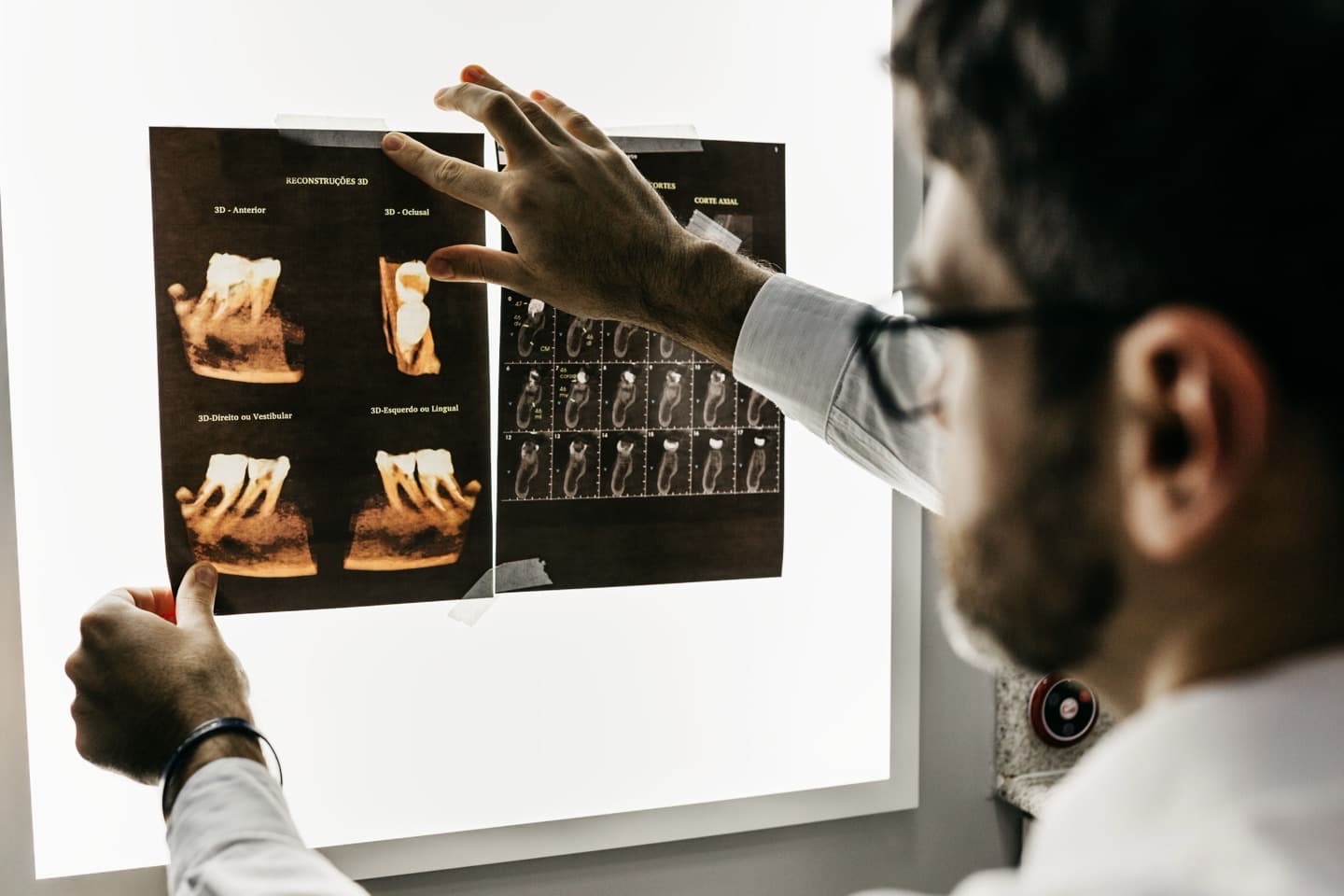
A panoramic X-ray is an imaging method that shows the upper and lower jaw, all teeth, and the surrounding bone structure in a single digital image. The machine rotates slowly around your head and captures this wide-angle image within a few seconds.
This image gives a general picture of many structures, including decay, gum-related bone loss, impacted teeth, and the jaw joint. But a panoramic X-ray is a "screening" tool by nature — when fine detail on a single tooth is needed, complementary imaging such as a small periapical X-ray of that specific tooth may also be used.
The vast majority of machines in use today are digital. Depending on the unit type, the measured effective dose ranges from 8.9 to 37.8 microsieverts (µSv) — I explain what that means in more detail in the radiation-safety section below (PMC, "Effective Dose From Direct and Indirect Digital Panoramic Units," retrieved 2026-08-24).
When Is a Panoramic X-Ray Requested During an Exam?
A panoramic X-ray isn't requested routinely at every exam; your dentist requests this imaging when there's a clinical reason to. The most common situations include:
- Evaluating impacted teeth — especially to see the position of wisdom teeth and their relationship to neighboring teeth
- Pain, clicking, or restricted movement in the jaw joint (TMJ)
- Suspicion of a cyst, abscess, or other lesion
- Assessing bone structure before an implant, extraction, or comprehensive treatment planning
- A first comprehensive exam, to get a general screening view of structures that can't be seen by eye
Do These Findings Always Come With a Symptom?
No, often they don't. In a study published on PMC, panoramic X-rays taken in patients with no symptoms showed at least one incidental (unexpected) finding in about 32% of cases; the most common was impacted teeth (PMC, "Incidental Findings in a Consecutive Series of Digital Panoramic Radiographs," retrieved 2026-08-24).
This finding shows why a panoramic X-ray is valuable not only when there's a symptom, but also as a general screening tool — though that doesn't mean it should be taken automatically at every exam; the decision still depends on the dentist's clinical assessment.
How Much Radiation Does a Panoramic X-Ray Involve, and Is It Safe?
When X-rays come up, the first question people usually have is about radiation safety. According to the American Dental Association (ADA), dental imaging is always planned according to the ALARA principle — meaning necessary exposures are kept "as low as reasonably achievable" (ADA, "X-Rays/Radiographs," retrieved 2026-08-24).
The numbers back this up: according to the ADA, dental imaging as a whole accounts for less than 1% of the total annual radiation dose from all medical imaging (ADA, same source). Measurements on digital panoramic units also show the effective dose staying in a low range — 8.9-37.8 microsieverts, depending on the unit type (PMC, same source).
Using digital sensors reduces this dose even further. A study comparing film and digital systems in general dental practices found that patient dose with digital phosphor-plate systems was reduced to about half compared to film, and exposure time with digital sensor (CCD) systems was cut roughly fourfold (PubMed, "Intraoral Radiology in General Dental Practices — A Comparison of Digital and Film-Based X-ray Systems," retrieved 2026-08-24).
As for frequency: the ADA states this varies from person to person and that there's no "one size fits all" — it depends on factors like age, current oral health, disease risk, and clinical findings, as determined by the dentist (ADA, same source). Modern digital equipment and beam collimation — directing the beam only at the area that needs it — are the main mechanisms behind this low dose.
What Is Intraoral Digital Scanning, and How Does It Work?
Intraoral digital scanning captures a three-dimensional digital image of the teeth and gums using a small handheld device. As the scanning tip moves through the mouth, the images are stitched together in real time on screen to build a 3D model.
This is the key difference from an X-ray: a panoramic X-ray images bone and internal tooth structure, while a digital scan captures only the visible surface — the shape — of the teeth and gums. Because they provide different types of information, one doesn't replace the other; they can be used together as complementary tools when needed.
Since no traditional impression material is needed, issues like the gag reflex or an unpleasant taste from holding impression material in the mouth are eliminated as well.
Which Treatments Use Digital Scanning?
Intraoral digital scanning is used as a supporting tool in planning many treatments that require a precise fit in the mouth, such as dentures, crowns, veneers, and clear aligners. It also plays an important role in preparing pre-treatment simulations during the smile design process — I covered that process in more detail in "How Does the Smile Design Process Work?"
Which treatment calls for digital scanning is determined by the dentist based on the treatment need; it isn't automatically requested for every intraoral procedure.
What Are the Advantages of Digital Scanning Over Traditional Impressions?
The most concrete advantage of digital scanning is accuracy. A systematic review and meta-analysis on the topic found that digital scanners produced a mean deviation of 137.86 microns, compared to 182.51 microns for traditional impressions (p<0.001) — meaning digital scanning is statistically significantly more accurate. The same study notes that 200 microns is generally used as the clinically acceptable threshold for prosthesis fit, and most digital scanner systems stay under that threshold (PMC, "Linear Accuracy of Intraoral Scanners for Full-Arch Impressions of Implant-Supported Prostheses: A Systematic Review and Meta-Analysis," retrieved 2026-08-24).
Alongside accuracy, comfort makes a real difference too: if the scan of one area doesn't come out well, only that area needs to be rescanned instead of repeating the entire impression from scratch.
| Traditional Impression | Intraoral Digital Scan | |
|---|---|---|
| Mean deviation (accuracy) | 182.51 microns | 137.86 microns |
| Impression material held in mouth | Yes | No |
| If an area needs to be redone | Entire impression is repeated | Only that area is rescanned |
Are These Methods Requested at Every Exam?
No. Neither a panoramic X-ray nor a digital scan is automatically requested at every check-up. In a routine check-up, if you have no symptoms and your previous imaging is recent, your dentist may proceed with a visual and manual exam alone.
These imaging methods come into play when there's a new symptom, a first comprehensive evaluation, a specific treatment plan, or a periodic check that calls for it. The decision is always made by the dentist based on that day's exam findings.
Are These Methods Safe During Pregnancy or for Children?
According to the UK's National Health Service (NHS), dental X-rays are considered safe during pregnancy and can be taken when needed for diagnosis or treatment; the X-ray beam doesn't point at the developing baby (NHS, Guy's and St Thomas' NHS Foundation Trust, "Dental X-rays," retrieved 2026-08-24). Still, letting your dentist know if you are or may be pregnant matters so the imaging can be planned appropriately.
Intraoral digital scanning doesn't involve X-rays, so it carries no additional restriction in this regard. For children, both methods can be used when needed, adjusted to age and clinical need; that decision is also made by the dentist during the exam.
Where Do These Imaging Methods Fit Into a Treatment Plan?
A panoramic X-ray and an intraoral digital scan don't replace a visual and manual exam — they complement it. One shows bone and internal tooth structure, the other shows the surface shape of the teeth and gums; together they help the dentist reach a more complete picture.
If your dentist recommends one of these methods at your next exam, it's usually because of a specific need tied to that day's findings or a planned treatment. If you have questions, you can ask your dentist directly during the exam and learn the reasoning behind the decision.
Frequently Asked Questions
Are a panoramic X-ray and an intraoral digital scan the same thing?
No. A panoramic X-ray uses radiation to show bone and internal tooth structure, while an intraoral digital scan captures only the surface shape of the teeth and gums without any radiation. Since they provide different types of information, one doesn't replace the other.
Is a panoramic X-ray taken at every exam?
No, it's only requested when the dentist sees a clinical reason for it; how often it's taken depends on the person's oral health.
Is a panoramic X-ray painful?
No, the procedure is painless and usually takes just a few seconds.
Can I get a panoramic X-ray while pregnant?
According to the NHS, dental X-rays are considered safe during pregnancy, but letting your dentist know beforehand matters so the imaging can be planned appropriately.
Is an intraoral digital scan painful or uncomfortable?
No, it's generally more comfortable than a traditional impression; it doesn't require impression material to sit in your mouth.
Is a digital scan automatically done before every treatment?
No, the dentist decides whether it's needed based on the treatment plan.
Can these methods be used with children?
Yes, they can be used when needed, adjusted to the child's age; that decision is made by the dentist during the exam.
When are panoramic X-ray results reviewed?
Thanks to digital imaging, results are usually reviewed the same day, during the exam.
Sources
- American Dental Association (ADA) — X-Rays/Radiographs
- PMC (National Library of Medicine) — Effective Dose From Direct and Indirect Digital Panoramic Units
- PMC — Incidental Findings in a Consecutive Series of Digital Panoramic Radiographs
- PubMed — Intraoral Radiology in General Dental Practices: A Comparison of Digital and Film-Based X-ray Systems
- PMC — Linear Accuracy of Intraoral Scanners for Full-Arch Impressions of Implant-Supported Prostheses: A Systematic Review and Meta-Analysis
- NHS (Guy's and St Thomas' NHS Foundation Trust) — Dental X-rays